 FR
FR AR
AR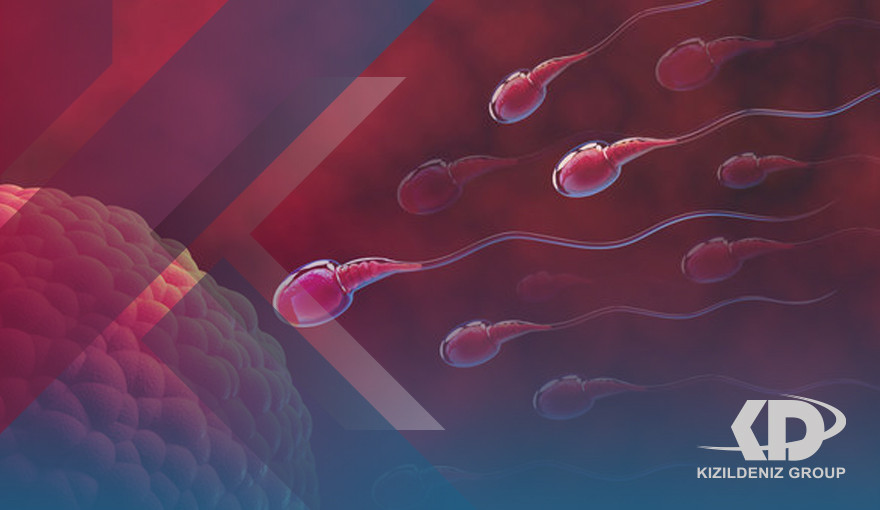
What is the specialty of Histology and Embryology:
Histology science, also known as microanatomy or microanatomy, is a branch of biology that studies the microanatomy of biological tissues. Tissue Science is the microscopic counterpart of total anatomy, which looks at larger visible structures without a microscope. Although one can divide microanatomy into organology, organ study, tissue science, tissue study, cell science, cell study, and modern use place all these subjects under the field of tissue science. In medicine, tissue pathology is a branch of tissue science that includes microscopic identification and the study of diseased tissue. In the field of paleontology, ancient biology refers to the tissue of fossil organisms.
History of Histology and Embryology Specialization:
In the 17th century, the Italian Marcello Malpighi used the microscope to study small biological entities. Some consider him the founder of the fields of tissue science and microscopy. Malbegi analyzed several parts of bat organs, frogs, and other animals under the microscope. During the study of lung structure, Malbegi observed membrane aerobic follicles and hair-like connections between the veins and the arteries, which he called blood capillaries. His discovery proved how the inhaled oxygen enters the bloodstream and serves the body.
In the 19th-century tissue science was an academic discipline in itself. The French anatomist Xavier Bichat introduced the concept of tissue in anatomy in 1801, and the term "tissue science" (German: Histologie), designed to refer to the "study of tissue," first appeared in a book by Carl Meyer in 1819. Twenty-one human tissues can be classified into the four categories currently accepted by tissue specialists. Jean Crovelier promoted the use of illustrations in tissues, which Bechat considered useless.
In the early 1830s, Burkini was invented with high accuracy.
During the 19th century, many stabilization techniques were developed by Adolph Hannover (chromate and chromic acid solutions), Franz Schulz, Max Schultz (nominal acid), Alexander Butlerov (formaldehyde), and Benedict Stelling (freezing).
Installation techniques were developed by Rudolf Heidenhain (1824-1898), who introduced Arabic glue. Salomon Stryker (1834-1898), who advocated a blend of wax and oil; and Andrew Pritchard (1804-1884) who in 1832 used a mixture of glue/glass. In the same year, Balsam Canada appeared on the scene, and in 1869 Edwin Klebs (1834-1913) reported that he had included his specimens in paraffin for several years.
The 1906 Nobel Prize in Physiology was awarded to tissue specialists Camilo Golgi and Santiago Ramón y Cajal. They had conflicting interpretations of the neural structure of the brain based on different interpretations of the same images. Ramon Y Kajal won the prize for his correct theory, and Golgi for the silver dye technique he invented to make it possible.
The importance of studying the specialty of Histology and Embryology:
For centuries, tissues have maintained their wonderful place in medical curricula. However, his teachings were influenced by new technological advances that reshaped the teaching of medicine into a more modern student-friendly model. Since its inception in the 18th century, tissue science has evolved alongside developments in microscopy and microscopy techniques, including immunochemistry. In the traditional curriculum of medical schools in the United States of America, especially after the First Flexner Report of 1910, tissue science was considered a very basic subject for a physician studying the "art and science" of medicine. In this era.
Teaching relied more on photometric microscopy and more or less on electron microscopy. However, at present, after the second Flexner report, which stressed the importance of integrating clinical subjects into the curriculum, the field is shifting towards using more electronic resources for teaching. These new resources rely on information techniques and electronic imaging methods that are more student-friendly, which have been effective and consistent in transmitting images, promoting self-learning, and being less expensive. In fact, in the last 25 years, most universities have relied on virtual microscopy with limited use of optical microscopes by students. This approach facilitated the integration of tissue curricula into pathological anatomy and provided an opportunity to promote self-learning and clinical relevance. In the age of competence-based curricula, the tissue remains essential and basic science indispensable in integrated units.
Histology and Embryology courses:
- Formation of jammies, implantation/general cellular structure, and epithelial tissue.
- Early development and differentiation between cells and tissues/types of connective tissue.
- Embryonic period (organ formation )/muscle system.
- Fetal period (functional development )/nervous system.
- Bone growth/skeletal tissue.
Fields of work for the Histology and Embryology major:
- Histology Technician
- Doctor
- Biologist
- Medical Laboratory Technician
Best Universities for Histology and Embryology in Turkey:
- Istanbul Medipol University